

Have you ever looked closely at someone’s eyes and noticed something striking? Maybe a vibrant ring of color circling their pupil, distinct from the rest of their iris? That eye-catching trait is what medical professionals call central heterochromia, and it’s far more fascinating than most people realize.
Central heterochromia occurs when the inner portion of your iris displays a different color than the outer ring. Instead of a single, uniform eye color, you have a two-toned iris that creates a distinctive appearance. While it might seem unusual, this condition is generally harmless and represents nothing more than a natural variation in how your body distributed pigment during development.
Many people don’t even realize they have this eye color pattern until someone points it out. Others spend their whole lives thinking they simply have hazel or green eyes with an interesting shade, never knowing there’s actually a medical term for what they’re seeing in the mirror.
This comprehensive guide explores everything you need to know about central heterochromia, from what causes it to develop, how to identify it properly, whether it requires medical attention, and what management options exist. Whether you have this condition yourself, you’re curious about a loved one’s eyes, or you’re just interested in the fascinating biology behind eye color variation, this article will provide answers to all your questions.
What Determines Eye Color?
Before we can understand central heterochromia, we need to understand how eye color works in the first place. The answer lies in a single substance: melanin.
Melanin is the pigment responsible for color in your skin, hair, and eyes. It’s the same pigment that gives your skin its tone and your hair its color. In your eyes, melanin deposits in the iris create the wide range of eye colors we see across the human population. More melanin means darker eyes (typically brown), while less melanin results in lighter eyes (blue, green, or gray).
But here’s where it gets interesting: the amount and distribution of melanin isn’t uniform across everyone’s irises. Some people have high concentrations everywhere, others have variations depending on where the melanin accumulated during fetal development. This is where our story of eye color variation truly begins.
The genetics of eye color involves multiple genes working together, not just one simple dominant-recessive relationship like we learned in basic biology class. The OCA2 gene, HERC2 gene, and many others influence how much melanin your body produces and where it deposits in your eyes.
During pregnancy, these genetic instructions guide melanin distribution. Most of the time, this happens evenly, creating a single, solid eye color. But sometimes, the distribution isn’t uniform. Melanin might concentrate more heavily in certain areas, creating distinctive patterns.
Light also plays a crucial role in how we perceive eye color. Light reflects off the melanin deposits and scatters through the eye tissue, creating the colors we observe. Different lighting conditions can make the same eyes appear different shades, which is why eye color can seem to change throughout the day.
Central Heterochromia: Definition and Visual Appearance
So what exactly is central heterochromia? Let’s define it clearly: central heterochromia is an eye condition where the inner ring around your pupil is a distinctly different color from the outer ring of your iris.
Think of it like a target or bullseye pattern. The pupil sits in the center (that’s the black circle that controls how much light enters your eye). Around this pupil is a ring of one color, which then transitions to a different color toward the edge of your iris. Unlike hazel eyes where colors blend gradually throughout the iris, central heterochromia features distinct, clearly defined color zones.
The visual appearance varies significantly from person to person. Some common color combinations include a golden or amber center with a blue outer ring, a brown center with green edges, or a yellow ring surrounded by hazel. The combinations are nearly endless because everyone’s melanin distribution is unique.
Many people with this eye color pattern never formally diagnose themselves. The condition might be so subtle in their case that they simply describe themselves as having hazel eyes or think their eye color is just unusual. Only when light hits their eyes at certain angles does the two-toned nature become obvious.
Others have high-contrast central heterochromia that’s immediately noticeable. A vibrant gold ring around the pupil transitioning to deep blue at the iris edge creates a striking appearance that people often comment on. These individuals are more likely to know they have central heterochromia because they’ve heard about it their whole lives.
The condition affects one or both eyes. Some people have it in just one eye while the other eye displays a single, solid color. Others have the pattern in both eyes, sometimes symmetrically and sometimes with different color combinations in each eye.
Types of Central Heterochromia
When we talk about iris color variation in general, medical professionals recognize several different types. Understanding these distinctions helps clarify what central heterochromia actually is and how it differs from similar-sounding conditions.
Complete heterochromia, also called heterochromia iridis when the variation is total, means each eye is completely a different color. One eye might be brown while the other is blue. This creates a striking appearance where your two eyes don’t match at all. Complete heterochromia is actually rarer than central heterochromia, affecting fewer than 200,000 Americans.
Sectoral heterochromia, sometimes called partial heterochromia, involves a wedge or slice of different color appearing on part of the iris. Imagine a slice of pie removed from the colored circle of your iris and replaced with a different color. This wedge pattern can be thin or take up a large portion of the iris. Sectoral heterochromia is characterized by a more irregular shape compared to the distinct ring pattern of central heterochromia.
Central heterochromia specifically means the color difference is concentrated around the pupil, creating that distinctive ring appearance. The color transition typically starts right at the pupil border and extends outward to varying degrees before transitioning to the outer iris color.
Heterochromia iridum and heterochromia iridis are simply alternative medical terms for the same general condition, with the distinction mainly in medical literature. Both refer to variation in iris color.
Understanding these distinctions matters because they can have different causes and different implications. Someone with acquired heterochromia that developed due to eye injury or medication might have any of these three types depending on which parts of the iris were affected.
Complete Heterochromia vs Central Heterochromia
The most common confusion is between complete heterochromia and central heterochromia. These are distinctly different conditions, and it’s important to understand how.
In complete heterochromia, your two eyes are fundamentally different colors. This affects the entire iris of each eye. One eye might have brown throughout, while the other is entirely blue. When you look in the mirror, you immediately notice the difference because your eyes are mismatched in the most obvious way possible.
Central heterochromia, on the other hand, affects only one iris—or technically both irises if you have it in both eyes, but the variation is within each iris, not between them. You still have only two eyes, obviously, but each eye contains two colors rather than your eyes being two different colors from each other.
Complete heterochromia is rarer, affecting fewer people in the general population. Because it’s more visually striking and unusual, it tends to be documented more often and is what most people think of when they hear the word “heterochromia” in casual conversation.
Central heterochromia is more subtle in many cases, which is why it often goes unnoticed or unnamed. You could have it your whole life without realizing there’s a specific term for your eye color pattern.
The causes can be different too. While both can be genetic, complete heterochromia might be more likely to be associated with certain genetic syndromes. Central heterochromia, being more subtle, is often purely a cosmetic variation with no underlying condition at all.
Sectoral Heterochromia vs Central Heterochromia
Sectoral heterochromia, the third major type of iris color variation, differs from central heterochromia in pattern and distribution.
With sectoral heterochromia, the different color appears as a wedge or sector—imagine a slice of your iris is a different color than the rest. This wedge can be thin like a hair or broad like a quarter of the iris, depending on the individual. The pattern is more irregular and asymmetrical compared to the distinct circular ring of central heterochromia.
The location of this sectoral color variation can be anywhere on the iris—top, bottom, left, right, or even multiple wedges. It doesn’t follow the concentric ring pattern that defines central heterochromia.
From a medical standpoint, sectoral heterochromia might have similar causes to central heterochromia, but the location of melanin variation is different. During fetal development, melanin distributed unevenly but concentrated in a specific sector rather than creating a central ring.
Both central and sectoral heterochromia are generally benign and don’t typically require treatment. Both can be congenital (present from birth) or acquired (developing later due to injury, inflammation, or medication). The distinction is mainly descriptive, helping eye care professionals communicate precisely about what they’re observing.
Some people have subtle sectoral heterochromia so mild that they think their eyes are just one solid color with slight variation. Others have dramatic wedges of different color that are immediately obvious.
Causes of Central Heterochromia
What causes this intriguing eye color variation? The answer depends on whether your central heterochromia is congenital (present from birth) or acquired (developed later).
The vast majority of central heterochromia cases are benign, meaning they’re not caused by any disease or medical condition. They simply represent normal human variation in how melanin distributed as your eyes developed. Your body’s melanin production and distribution are controlled by genetics, but the exact pattern is somewhat random and can vary even between identical twins.
Think of it this way: melanin distribution during fetal development is like paint being applied to a surface. Most of the time, it spreads evenly, but sometimes—for reasons we don’t fully understand—it concentrates more heavily in certain areas. When this happens to concentrate around the pupil, creating that ring pattern, you get central heterochromia.
This benign form of heterochromia doesn’t require any treatment or medical intervention. It doesn’t affect your vision, increase your risk of eye disease, or cause any health problems whatsoever. You can live your entire life with central heterochromia and never experience any negative consequences related to it.
However, central heterochromia can sometimes be a sign of an underlying medical condition. This acquired form develops after birth due to specific causes like eye injury, infection, inflammation, or medication side effects. When central heterochromia develops later in life, that’s the time to consult with an ophthalmologist to determine what’s causing the change.
Genetic Heterochromia and Central Heterochromia
Most cases of central heterochromia are genetic in origin, meaning they’re present at birth and caused by hereditary factors. But here’s an important clarification: genetic doesn’t necessarily mean inherited.
Genetic heterochromia develops because of genetic instructions—your DNA—but in most cases, it’s not passed down from parents to children. Instead, it’s caused by a random genetic variation that occurs during your fetal development. Your parents might not have heterochromia at all, yet you developed it due to this random variation.
This distinction confuses many people. Yes, heterochromia is genetic. No, you usually won’t inherit it from your parents. Genetic simply means it’s determined by your genes and DNA, while inherited means it came from your parents’ genetic material.
Some families do have central heterochromia running through multiple generations, suggesting a hereditary pattern. But these cases are relatively rare. Most people with central heterochromia developed it due to spontaneous genetic variation during their own fetal development.
The genes involved in eye color—primarily OCA2 and HERC2, along with many others—influence melanin production. A variation in how these genes are expressed can lead to uneven melanin distribution. During the critical weeks of fetal development when your eyes are forming, if melanin concentrates around the pupil more than elsewhere, you’ll develop central heterochromia.
Understanding this distinction is important if you have central heterochromia and are wondering whether your children will develop it. The answer is: probably not, unless they also experience the same random genetic variation. It’s not impossible, but it’s not the likely inheritance pattern for central heterochromia.
Acquired Heterochromia: When Central Heterochromia Develops Later in Life
While most central heterochromia is congenital, some people develop color variation in their irises later in life. This is called acquired heterochromia, and it has very different causes than congenital heterochromia.
Acquired central heterochromia means something happened to damage or affect the melanin-producing cells in your iris, causing a color change after you were born. This might be an injury, an infection, inflammation, medication effects, or a medical condition affecting your eyes or nervous system.
If you notice your iris color changing in adulthood, this is worth investigating with an eye care professional. While many causes of acquired heterochromia are benign, some might indicate an underlying health issue that needs attention.
Eye injury is a common cause of acquired heterochromia. Trauma to the eye can damage melanin-producing cells, causing color changes in the affected iris. The injury might occur from an accident, sports impact, or other trauma. The resulting color change might be immediate or develop gradually as the eye heals.
Infection in the eye can also cause acquired heterochromia. Certain infections, particularly those that cause inflammation of the iris (iritis) or the entire uveal tract (uveitis), can damage melanin distribution and cause color changes.
Inflammation of the iris, whether from infection or other causes, can alter how melanin is distributed. Conditions like Posner-Schlossman syndrome, characterized by intermittent anterior uveitis, can cause iris color changes.
Medical conditions affecting the eye, such as pigment dispersion syndrome where melanin granules shed from the iris, can cause heterochromia. Iris ectropion syndrome, where iris tissue extends abnormally, can also result in color changes.
The key difference between congenital and acquired heterochromia is the timing. If you’ve had the same eye color pattern your whole life and it hasn’t changed, it’s almost certainly congenital. If you notice a new color variation appearing or an existing pattern changing, that’s acquired heterochromia, and you should see an ophthalmologist to determine the cause.
Melanin Distribution: The Science Behind Central Heterochromia
Understanding the science of how your irises actually develop helps explain why central heterochromia happens in the first place.
Melanin, that crucial pigment we keep mentioning, is produced by cells called melanocytes. During fetal development, these melanocyte cells migrate into the iris and deposit melanin according to genetic instructions. In most people, this distribution happens relatively evenly, resulting in a single, solid eye color.
But the distribution process isn’t perfectly uniform across every person’s iris. Factors we don’t completely understand yet influence where exactly melanin concentrates. Some areas might receive more melanin deposits than others. When this happens preferentially around the pupil, creating a higher concentration of melanin in the inner ring than the outer ring, you get central heterochromia.
The science here is that the amount of melanin determines how light is absorbed and scattered. More melanin absorbs more light, making that area appear darker or warmer-toned. Less melanin allows more light to scatter, creating lighter or cooler-toned appearances. This is why the inner and outer rings of an iris with central heterochromia appear different—they have genuinely different melanin concentrations.
The timing of this melanin distribution happens during specific weeks of fetal development. Your eyes are forming and developing rapidly during the second and third trimesters of pregnancy. The exact weeks when melanin deposits heavily influence the final color and pattern you’ll have.
Once your iris development is complete and you’re born, the melanin distribution pattern is essentially permanent in the case of congenital heterochromia. You won’t develop more central heterochromia than you started with, and the pattern won’t change on its own.
This is why acquired heterochromia, which happens when something damages or changes existing melanin distribution, is different from congenital heterochromia. The latter is set during development. The former involves changes to an already-formed eye structure.
Is Central Heterochromia Genetic or Hereditary?
This question confuses many people, and it’s worth clarifying carefully because the answer involves understanding the difference between genetic and hereditary.
Central heterochromia is genetic, meaning it’s determined by your genes and DNA. Genetics refers to the role of your genetic material in determining your traits. If you have central heterochromia, it’s because your genes influenced melanin distribution in a particular way.
However, central heterochromia is usually not hereditary, meaning it doesn’t typically run in families or get passed from parents to children. Hereditary refers to traits inherited from your parents. While some families do show patterns of heterochromia across generations, this is relatively uncommon.
Most people with central heterochromia have parents without heterochromia. They developed the trait due to a random genetic variation that occurred during their own development, not due to inheriting a specific heterochromia-causing gene from their parents.
If you have central heterochromia and you’re wondering whether your children will develop it, the honest answer is: probably not, but it’s not impossible. The same random genetic variation that occurred in you during your fetal development could theoretically occur in your child during their fetal development. But this isn’t a guaranteed inheritance.
Genetic counselors sometimes use central heterochromia as an example when explaining the difference between genetic (determined by genes) and hereditary (inherited from parents). You can have a genetic condition without having inherited it from your family.
Some research suggests that certain patterns of heterochromia might have slightly increased likelihood in families where a parent has it, but the actual inheritance risk is low. Most genetic counselors don’t recommend genetic testing or worry about inheritance if you have central heterochromia and are planning to have children.
The bottom line: central heterochromia is a genetic trait in most cases, but you probably won’t pass it to your children, and your children probably won’t develop it unless they experience the same random genetic variation you did.
Medical Conditions Associated With Central Heterochromia
While most central heterochromia is benign and unrelated to any medical condition, some underlying diseases can cause heterochromia as a symptom. These conditions typically cause acquired heterochromia, developing after birth rather than being present from birth.
Knowing these conditions exists matters because if you develop heterochromia later in life, determining whether it’s related to one of these underlying issues could be important for your health.
Eye injury or trauma is probably the most common cause of acquired heterochromia. When the iris is damaged by injury, the melanin-producing cells might be disrupted, causing color changes. This could be from sports injuries, accidents, or other eye trauma.
Infection in the eye can damage the iris and alter melanin distribution. Serious eye infections can cause inflammation and structural changes that result in color variation. This is why sudden color changes in your iris warrant an eye exam.
Inflammation of the iris without infection can also cause heterochromia. Conditions causing iris inflammation need proper diagnosis and treatment, as chronic inflammation can affect your vision long-term.
Pigment dispersion syndrome involves melanin granules being shed from the iris into other parts of the eye. This can result in heterochromia as the iris loses pigment in areas. People with pigment dispersion syndrome have increased risk of developing glaucoma.
Iris ectropion syndrome, where iris tissue extends abnormally, can alter the appearance and potentially cause color changes or variation.
Several genetic syndromes can include heterochromia as one of their features. These include Horner syndrome, Waardenburg syndrome, Sturge-Weber syndrome, and neurofibromatosis. In these cases, heterochromia is one of multiple symptoms rather than an isolated finding.
Medications That Can Cause Central Heterochromia

Certain medications, particularly eye drops used to treat glaucoma, can cause changes in iris color over time. This is an important side effect to be aware of if you’re using these medications long-term.
Prostaglandin analog eye drops are the primary medications associated with iris color changes. These include latanoprost, travoprost, and bimatoprost. These drugs work by increasing fluid drainage from the eye, reducing eye pressure in glaucoma patients. A side effect of this drug class is increased melanin production in the iris.
Latanoprost, sold under brand names like Xalatan and Xelpros, can cause a gradual darkening or color change of the iris. Studies show that up to 33% of people taking latanoprost for 5 years or longer experience some degree of iris color change. The change tends to be gradual, happening over months or years rather than suddenly.
Travoprost, including brands like Travatan Z and Izba, has similar iris-coloring effects as latanoprost. The mechanism is the same—the medication stimulates melanin production in the iris, causing it to darken or change color over time.
Bimatoprost, available as Eyrieda, Lumigan, or Sturiban, can also cause iris color changes. Additionally, bimatoprost has another side effect that might be more noticeable to patients: it can stimulate eyelash growth and darken eyelashes.
Latisse is interesting because it was originally a glaucoma medication (formulated as Lumigan for that purpose) but is now primarily marketed as a cosmetic product to thicken and darken eyelashes. Because it’s the bimatoprost molecule, it carries the same potential for iris color changes, though when used as a topical eyelash application, the risk is lower than with eye drops applied directly to the eye.
If you’re using prostaglandin analog eye drops, it’s worth knowing about this potential side effect, though it’s generally not dangerous or harmful to your vision. The color changes are usually permanent or very slow to reverse even after stopping the medication. The important thing is being informed and monitoring your eyes.
Central Heterochromia and Underlying Eye Disease
Central heterochromia itself doesn’t cause eye disease or vision problems. The condition is purely cosmetic in most benign cases. However, if heterochromia develops acquired, it might be a sign of an underlying eye condition that does need attention.
Glaucoma can sometimes present with iris changes, including heterochromia. While heterochromia alone isn’t a glaucoma symptom, if you develop heterochromia and have other glaucoma risk factors, an eye exam to check your eye pressure would be wise.
Uveitis, inflammation of the uveal tract (which includes the iris), can cause heterochromia. Uveitis can damage the structures of your eye and affect vision if left untreated. It requires proper diagnosis and treatment.
Iris cysts or tumors, though rare, can potentially affect iris appearance and cause heterochromia. This is another reason sudden iris color changes warrant professional evaluation.
Angle closure glaucoma, a medical emergency, can cause iris changes and heterochromia as part of the acute attack. But this would come with other obvious symptoms like severe pain and vision changes.
Most people with central heterochromia do not have any underlying eye disease. But the timing of when it develops matters. Congenital heterochromia is essentially never associated with disease. Acquired heterochromia—developing after birth—warrants investigation to determine its cause.
How to Identify Central Heterochromia
Identifying central heterochromia is actually quite straightforward: look closely at your irises in good lighting.
Central heterochromia appears as a distinct ring of different color around your pupil. The pupil is the black circle in the center of your eye that opens and closes to control light. Right around this black circle, you might see a ring of color—perhaps gold, amber, brown, green, or another shade—that’s distinctly different from the color of the rest of your iris.
The key visual distinction is that the inner and outer rings have a fairly clear color boundary. It’s not a gradual blending of colors like you’d see in hazel eyes. Instead, there’s a noticeable transition from the inner ring color to the outer iris color.
Some people have subtle central heterochromia where the color difference is only visible in certain lighting conditions or when looking very closely. Others have dramatic, high-contrast central heterochromia that’s immediately obvious to anyone looking at them.
The best way to check if you have central heterochromia is to look in a mirror in bright, natural lighting. Position yourself near a window or outside if possible. Look closely at your iris, focusing on the area immediately surrounding your pupil. Ask yourself: is the color here different from the rest of my iris?
You might also take a close-up photograph of your eye in good lighting. Sometimes the camera picks up details that are hard to see in the mirror. Compare the inner ring color to the outer ring color in the photo.
Some people never notice they have central heterochromia until someone else points it out or until they read about it somewhere. Others have known their whole life that their eyes look different but never had a name for it. Both scenarios are common.
Keep in mind that what you might perceive as central heterochromia could also be hazel eyes, which are simply a blended mixture of colors throughout the iris. The distinction is whether you have a clear ring pattern (central heterochromia) versus blended colors throughout (hazel).
Central Heterochromia vs Hazel Eyes: What’s the Difference?
This is probably the most common source of confusion about central heterochromia. People often mistake hazel eyes for central heterochromia and vice versa. Let’s clarify the distinction.
Hazel eyes are characterized by a mixture or blend of colors throughout the iris. Brown, green, gold, and amber tones intermix across the entire iris surface. The colors appear to blend together and shift depending on the lighting. In different light, hazel eyes might appear more green, more brown, or more golden as the light reflects differently off the mixed colors.
Central heterochromia has distinct, separate color zones. There’s a clear inner ring of one color and an outer ring of a different color. The transition between these zones is relatively sharp, not gradually blended.
Hazel eyes create an appearance where colors are distributed throughout, while central heterochromia creates an appearance where colors are organized in concentric rings.
The difference matters because they’re fundamentally different eye color patterns. While both can look beautiful and striking, they’re distinct conditions. If someone tells you they have hazel eyes, you know the colors are blended. If they have central heterochromia, you know the colors are organized in rings.
Some people genuinely have both features—perhaps they have central heterochromia but the colors blend slightly at the boundary, or their hazel coloring includes a hint of the ring pattern. Eyes are complex and individual variation is enormous.
The easiest way to distinguish them is to look closely in good lighting and ask yourself: are the colors blended together throughout (hazel) or organized in distinct rings (central heterochromia)? With careful observation, the difference becomes clear.
Many people with subtle central heterochromia have lived their whole lives thinking they simply have hazel or green or blue eyes without realizing their eyes actually have the two-toned ring pattern characteristic of central heterochromia.
Color Combinations in Central Heterochromia
Central heterochromia appears in countless color combinations because everyone’s melanin distribution is unique. Let’s explore some of the most common patterns people report.
Blue outer iris with golden center is a classic combination. The contrast between cool blue tones and warm golden tones creates a striking appearance. This pattern often appears in people of European descent with naturally lighter eye colors.
Green outer iris with amber or brown center is another common pattern. Green eyes are relatively uncommon in the general population, and when combined with central heterochromia, the effect is quite distinctive. The warm center contrasts beautifully with the cooler green exterior.
Brown outer iris with golden center appears in people with more melanin overall. The brown provides a darker outer ring, with a noticeably lighter golden or amber ring immediately around the pupil.
Hazel outer iris with brown center represents a more subtle version of central heterochromia. The distinction between outer and inner colors is less dramatic than other combinations but still noticeable upon close examination.
Gray outer iris with green or blue center is relatively rare but strikingly beautiful when it occurs. The cool gray tones in the outer ring contrast with warmer inner colors.
The specific color combination you have depends on how much melanin is in each ring and how light reflects through the iris tissue at different depths. Deeper structures in the iris might contain different amounts of melanin than the surface layers.
It’s worth noting that the apparent colors can shift slightly with lighting and with what you’re wearing. A ring that appears golden in daylight might look more amber in indoor lighting. What appears green in certain light might look blue in different conditions. This is normal and doesn’t mean your eye color is actually changing, just that light is reflecting and scattering differently.
Can Central Heterochromia Appear in Both Eyes?
Yes, central heterochromia can appear in one eye or both eyes, depending on how melanin distributed during your fetal development.
Having central heterochromia in only one eye is relatively common. Your left eye might have a golden ring while your right eye displays a single, solid color. This asymmetry is perfectly normal and doesn’t indicate any problem.
Having central heterochromia in both eyes is also common. In this case, both your irises display the ring pattern. Sometimes the color combinations are identical in both eyes, sometimes they differ. You might have blue outer/gold inner in both eyes, or you might have blue outer/gold inner on the left side and green outer/brown inner on the right side.
The specific pattern reflects how melanin happened to distribute during your individual fetal development. It’s unique to you, just like your fingerprints or your DNA.
Having bilateral central heterochromia (in both eyes) might make the condition more noticeable since the pattern appears in both irises. But it’s no different from a medical or health perspective than having it in only one eye. Either way, it’s a benign cosmetic variation.
Some people find their bilateral central heterochromia to be one of their most distinctive physical features. Others have such subtle bilateral heterochromia that they don’t notice it about themselves until someone else mentions it.
The development of central heterochromia in one eye versus both eyes is determined entirely by genetics and random variation in melanin distribution during fetal development. There’s no medical reason to prefer one pattern over another.
Diagnosis of Central Heterochromia
Diagnosing central heterochromia is remarkably straightforward because it’s primarily a visual observation. An eye care professional doesn’t need special testing or imaging to diagnose central heterochromia. They simply look at your iris.
An ophthalmologist or optometrist examining your eyes will use a slit lamp—a specialized microscope with a bright light designed to examine the eye in detail. Under magnification with the slit lamp, the ring pattern of central heterochromia becomes very clear. The different colors and their boundaries are easy to see.
Your eye care professional will ask about your medical history, including whether you’ve always had this eye color pattern (suggesting congenital heterochromia) or whether it developed recently (suggesting acquired heterochromia). They’ll ask whether anyone in your family has heterochromia.
If the heterochromia is congenital and stable, that’s essentially the entire diagnosis. An ophthalmologist confirms you have central heterochromia and reassures you that it’s benign and requires no treatment.
If the heterochromia is acquired or if there’s any concern about underlying conditions, further testing might be warranted. This could include eye pressure measurements if glaucoma is a concern, special imaging to examine the eye structures, or blood tests if a systemic condition is suspected.
Photography is often used in the diagnostic process. Your eye care professional might photograph your iris to document the heterochromia, create a record for comparison if they want to monitor for changes, and sometimes to share with you so you can see the pattern clearly yourself.
Most people with central heterochromia never need special diagnostic testing. The visual diagnosis combined with a normal eye exam is sufficient. The diagnosis is that you have central heterochromia, it’s benign, and no treatment is necessary.
When to See an Ophthalmologist for Central Heterochromia
Most people with central heterochromia never need to see a doctor specifically about the heterochromia itself. But there are situations when consulting an eye care professional makes sense.
You should see an ophthalmologist if you notice a sudden change in your iris color. If your eyes have been one color your whole life and you suddenly notice different coloring or a new ring pattern developing, that’s acquired heterochromia and warrants professional evaluation. Something caused that change, and determining what is important.
Any of the following symptoms accompanying iris color changes warrant prompt evaluation: eye pain, vision changes, redness, discharge, sensitivity to light, or a feeling of something in your eye. These suggest inflammation or infection rather than simple cosmetic heterochromia.
If you develop heterochromia and have personal or family history of eye disease, a thorough evaluation is wise. Certain conditions that cause acquired heterochromia also increase risk of vision problems.
If you have congenital heterochromia and you’re having regular eye exams for general health (which everyone should do), your eye care professional will note the heterochromia and monitor for any changes. Regular eye exams are important for everyone regardless of heterochromia.
If you have a genetic syndrome that includes heterochromia as one of its features—like Horner syndrome, Waardenburg syndrome, or Sturge-Weber—you should maintain regular ophthalmologic care because these syndromes carry risks for other eye complications.
If you’re using prostaglandin analog eye drops for glaucoma, you should be aware that iris color changes are a potential side effect. Discuss this with your ophthalmologist if you’re concerned about the changes.
Reassuringly, most people with simple, stable, benign central heterochromia don’t need any special ophthalmologic care beyond the regular eye exams everyone should get. But these situations above represent times when professional evaluation is warranted.
Is Central Heterochromia Harmful or Dangerous?
This is the question that brings many people to research central heterochromia in the first place. People worry: is this eye color variation a sign of something serious? Will it affect my vision? Is it dangerous?
The straightforward answer for congenital central heterochromia is: no, it’s not harmful or dangerous. Central heterochromia itself is purely cosmetic. It doesn’t affect your vision, it doesn’t increase your risk of eye disease, it doesn’t cause any health problems. You can live your entire life with central heterochromia without it ever affecting your health or wellbeing.
The condition doesn’t progress or worsen over time. You won’t gradually lose vision because of central heterochromia. You won’t develop other eye problems because of it. The heterochromia you have now is essentially the heterochromia you’ll have for life (in congenital cases).
Some people worry that central heterochromia indicates an underlying medical condition. While acquired heterochromia (developing after birth) can sometimes indicate an underlying issue, congenital heterochromia almost never does. If you’ve had the same eye color pattern your entire life, it’s almost certainly benign.
The only situations where central heterochromia might warrant concern are if it’s part of a genetic syndrome that has other health implications. For example, Waardenburg syndrome includes heterochromia as one feature but also includes potential hearing loss and other considerations. If you have central heterochromia as part of a broader syndromic pattern, management of the overall syndrome is what matters.
For the vast majority of people with central heterochromia, it’s simply an interesting variation in eye color. It’s not harmful, it’s not dangerous, and it requires no medical intervention. The biggest “problem” most people face is simply explaining what they have when someone asks about their unusual eyes.
Treatment for Central Heterochromia
Here’s the simple truth: central heterochromia doesn’t require treatment because it’s not a medical condition that needs treating. There’s no medication, surgery, or therapy that can change the heterochromia itself.
Since central heterochromia is purely cosmetic in benign cases, treatment isn’t medically necessary. You don’t need to do anything about it from a health perspective.
However, if you’re interested in changing how your eyes appear for cosmetic reasons, you do have options.
Colored contact lenses are the primary cosmetic option for central heterochromia. If you want to hide the heterochromia and have uniform eye color instead, you can wear colored contacts. These work just like regular contact lenses but are tinted to provide color. You can choose any color you prefer, completely obscuring the heterochromia.
Colored contacts are widely available, relatively affordable, and safe when used properly. You do need a contact lens prescription and training on how to insert, remove, and care for them. Your eye care professional can provide guidance on this.
Some people with central heterochromia choose colored contacts to achieve a dramatic look—perhaps choosing a color completely different from their natural heterochromatic eyes. Others simply want uniform color and choose contacts that closely match their natural iris color.
If your central heterochromia developed due to an underlying medical condition, treating that underlying condition is important. For example, if medication caused the color change, discussing alternatives with your physician might be warranted. If inflammation caused it, treating the inflammation is necessary. But the heterochromia itself wouldn’t be directly treated; rather, you’d be treating whatever caused it.
Most people with central heterochromia choose not to treat or hide it. Instead, they accept it as part of their unique appearance. Many people find their central heterochromia to be one of their most distinctive and attractive features.
Management and Living With Central Heterochromia
If you have central heterochromia, managing it is remarkably simple: accept it as part of who you are and take normal eye care measures to protect your vision.
Eye protection and eye health maintenance apply to everyone. Wearing sunglasses with UV protection shields your eyes from sun damage. Protecting your eyes during activities with injury risk prevents trauma that could potentially cause acquired heterochromia or other problems.
If you use a computer regularly, taking breaks and using proper screen positioning helps prevent digital eye strain. Keeping your eyes moist, particularly if you spend long periods in dry environments or using devices, maintains eye comfort.
Regular eye exams help monitor your overall eye health. Even though central heterochromia doesn’t require specific monitoring, regular eye exams catch other potential eye conditions early.
If you’re using prostaglandin analog eye drops for glaucoma, regular follow-up with your ophthalmologist ensures your glaucoma is controlled and allows monitoring for medication side effects including iris color changes.
Some people with central heterochromia mention that they’ve become quite attached to their distinctive eyes. They use them as their identifying feature, incorporating them into how they present themselves to the world. Others barely think about their heterochromia, viewing it as simply how their eyes happen to look.
If you have social anxiety about your appearance or if your heterochromia is causing you psychological distress, talking with a counselor or therapist can help you work through those feelings. Most people eventually embrace their unique eye color as part of their identity.
Photography enthusiasts with central heterochromia often discover they love capturing close-up photos of their eyes. The ring pattern photographs beautifully and makes for striking images. Your distinctive eyes can become an asset in self-portraits and photography projects.
Central Heterochromia in Different Ethnicities
Central heterochromia appears across all ethnic and racial groups. However, some patterns and prevalence rates might differ based on genetics and melanin distribution variations that differ across populations.
Lighter eye colors, including blue, green, and gray, are most common in European-descended populations. Because central heterochromia creates visible color variation, it’s more noticeable in people with lighter eye colors where the contrast between the inner and outer rings is more dramatic. A golden center is more visually distinct against blue than it might be against brown.
This doesn’t mean central heterochromia is more common in European-descended populations; it might simply be more noticeable because lighter eye colors create more contrast. The underlying genetic variation in melanin distribution probably occurs at similar rates across populations.
Darker eye colors, more common in African, Asian, and Mediterranean-descended populations, can also display central heterochromia. The pattern might be less visually obvious than in lighter-eyed individuals, but it’s still present. A subtle brown-on-brown ring pattern requires careful observation to detect, but it’s there.
This is why central heterochromia is sometimes described as underdiagnosed. Many people with subtle heterochromia in darker eye colors never realize they have it because the contrast is minimal.
Research on the actual prevalence of central heterochromia across different ethnic groups is limited. Most studies focus on complete heterochromia, which is more striking and therefore more likely to be documented and studied.
What we do know is that central heterochromia can appear in anyone, regardless of ethnic or genetic background. The fundamental biology—melanin production and distribution during fetal development—applies universally across all human populations.
Central Heterochromia in Children vs Adults
Children with congenital central heterochromia have had the condition since birth, and it becomes more noticeable as their personality and appearance develop. Young children might not be particularly aware of their heterochromia or its uniqueness.
As children grow into their school years, they might become more aware that their eyes look different from other kids’ eyes. This can spark curiosity in both the child and their peers. Some children handle this with confidence, explaining their unique eye color to curious classmates. Others might develop self-consciousness about their appearance.
Parents of children with central heterochromia can help by normalizing the difference and teaching their child that it’s a unique, interesting feature rather than something wrong or bad. Helping them understand the biology behind why it happens can foster appreciation for their distinctive eyes.
Teenagers with central heterochromia might experience increased self-consciousness about their appearance as they become more aware of physical differences. Supporting teens in accepting and even celebrating their unique features helps build confidence during these important developmental years.
Adults with central heterochromia have usually had years to adapt to their distinctive eyes. Many adults report that by the time they reach adulthood, they’ve incorporated their heterochromia into their self-image and identity. Some mention that they’ve received compliments about their eyes throughout their lives, which reinforces positive feelings about this feature.
Older adults with congenital heterochromia continue to have the same eye color pattern they’ve had for their entire lives, though aging and presbyopia (age-related vision changes) might affect other aspects of their eye health.
Adults who develop acquired heterochromia face different psychological and practical considerations since they’re experiencing a change in their appearance. The novelty and potential medical concern about what caused the change adds complexity compared to people who’ve always had heterochromia.
Congenital Central Heterochromia: Present From Birth
Congenital central heterochromia means you’re born with the condition. You develop it during fetal development, and it’s present from birth (or becomes apparent shortly after birth as your eye color fully develops).
Newborns often have blue eyes regardless of their genetic heritage because of how light interacts with the iris at birth. The full iris color typically develops within the first few months to a year of life as melanin deposition continues.
If you have congenital central heterochromia, the ring pattern might not be immediately obvious at birth due to the general blue eye appearance all newborns have. But as melanin deposits and your true eye color develops, the heterochromia pattern becomes apparent.
Parents sometimes notice the distinctive ring pattern in their baby’s eyes during their first year of life. A pediatrician or eye care professional can confirm that it’s central heterochromia.
Congenital central heterochromia is stable and permanent. Once you’re born with it, the pattern doesn’t change throughout your life. You’ll have the same eye color pattern at age 5, age 25, age 65, and beyond.
This permanence is actually reassuring. If you’ve had the same eye color your whole life, you know it’s benign. There’s no worry that it will suddenly change or indicate a developing medical problem.
The vast majority of congenital central heterochromia is purely benign with no underlying medical condition. It’s simply a cosmetic variation in how melanin distributed during your fetal development.
Some genetic syndromes can include congenital central heterochromia as one of their features, but this is relatively rare. If you have congenital heterochromia and a diagnosed genetic syndrome, your medical care focuses on managing the syndrome as a whole.
Acquired Central Heterochromia: Development After Birth
Acquired central heterochromia develops after birth due to specific causes affecting your iris. This is different from congenital heterochromia in important ways.
If you notice your iris color changing or a new ring pattern developing, that’s acquired heterochromia. The change might be sudden or gradual depending on what caused it.
Eye injury is the most common cause of acquired heterochromia. Trauma to your eye can damage melanin-producing cells or alter the iris structure, causing color changes. This could result from an accident, sports injury, or other eye trauma. The color change might occur immediately after the injury or develop gradually as the eye heals.
Medication side effects, particularly from prostaglandin analog eye drops used for glaucoma, can cause gradual iris color darkening or changes. These medications stimulate melanin production, leading to increased pigmentation over months or years of use.
Inflammation of the iris, from infection or other causes, can alter melanin distribution and cause heterochromia. Conditions like uveitis causing iris inflammation need proper treatment, both to address the inflammation and to prevent vision complications.
Other medical conditions affecting eye structures can potentially cause acquired heterochromia. This is why any sudden iris color change warrants professional evaluation.
The key difference between acquired and congenital heterochromia is the timing. Something specifically caused the change, and determining what that something is might be medically important.
Acquired heterochromia isn’t necessarily concerning, but it warrants investigation. Your eye care professional can help determine the cause and whether any treatment is necessary.
For the latest, updated, and unique information, visit Perfect Order Card.
Central Heterochromia and Vision: Does It Affect Your Eyesight?
No, central heterochromia does not affect your vision. Your eyesight and visual acuity are completely unrelated to whether you have heterochromia.
The iris and the melanin distribution within it are purely about eye color. The structures actually responsible for vision—the lens, retina, optic nerve, and visual cortex—function normally in people with central heterochromia.
Melanin in the iris serves to protect the eye from excess light and helps control light entering the eye through pupil dilation and constriction. The amount of melanin doesn’t significantly affect vision as long as the pupil can still function normally.
People with central heterochromia see the world exactly the way people without heterochromia do. They have the same visual acuity, color vision, night vision, and overall eyesight as anyone else.
Some people worry that because their iris looks different, something must be different about how they see. This is understandable but unfounded. Central heterochromia is purely cosmetic. The only difference it makes to your eyes is visual appearance.
If someone with central heterochromia experiences vision problems, those problems are unrelated to the heterochromia. They might be due to unrelated eye conditions like myopia, hyperopia, astigmatism, presbyopia, or other vision issues that occur in people with or without heterochromia.
This is an important reassurance for people with central heterochromia: your distinctive eye color has zero impact on how well you see the world.
Myths and Misconceptions About Central Heterochromia
Several myths and misconceptions about central heterochromia circulate online and in popular culture. Let’s address the most common ones.
Myth: Central heterochromia means you’re a supernatural being or have special powers. Reality: It’s simply a variation in melanin distribution. No supernatural elements involved, no special powers granted.
Myth: Central heterochromia is dangerous and requires immediate medical attention. Reality: Congenital central heterochromia is benign and requires no medical treatment. Acquired heterochromia warrants evaluation, but the heterochromia itself isn’t dangerous.
Myth: Central heterochromia is caused by disease or medical conditions. Reality: Most cases are congenital and unrelated to any disease. Some acquired cases might indicate an underlying issue, but this is the exception, not the rule.
Myth: Central heterochromia will get worse or change over time. Reality: Congenital heterochromia is stable and doesn’t progress. It stays the same throughout your life.
Myth: Central heterochromia is so rare that if you have it, something must be very wrong. Reality: While technically not extremely common, central heterochromia is common enough that many people have it. It’s probably underreported because people don’t always realize they have it or don’t know the medical term.
Myth: You’ll definitely pass central heterochromia to your children. Reality: Most people with central heterochromia didn’t inherit it, and most won’t pass it to their children. Inheritance is possible but not the typical pattern.
Myth: Central heterochromia affects intelligence, personality, or ability. Reality: Eye color and iris patterns have nothing to do with cognitive abilities, personality traits, or capabilities.
Myth: Central heterochromia is a sign of a rare genetic condition. Reality: Most congenital central heterochromia is simply a cosmetic variation without any associated genetic syndrome.
Myth: Contact lenses are unsafe if you have central heterochromia. Reality: Contact lenses are safe and can be worn by anyone with a valid prescription, including people with central heterochromia.
Myth: Central heterochromia will damage your vision over time. Reality: It won’t. Your vision will remain unchanged regardless of your heterochromia.
Central Heterochromia in Animals vs Humans
Central heterochromia appears much more commonly in animals than in humans, which is a fascinating biological pattern.
Dogs frequently display heterochromia of various types. Certain dog breeds like Siberian Huskies are particularly known for heterochromia. Some dogs have complete heterochromia with each eye completely different colors, while others display patterns more like central or sectoral heterochromia. The distinctive eye patterns in dogs are sometimes even selected for in breeding.
Cats, particularly white cats, commonly have heterochromia. This is sometimes linked to the genes that produce white coat color. Complete heterochromia in cats (one blue eye and one orange or green eye) is well-documented and even celebrated in cat breeding communities.
Horses display heterochromia regularly. Various patterns appear in horse populations, and different colors in the eyes don’t particularly concern equine veterinarians.
Other animals including cattle, some bird species, and other mammals display heterochromia with varying frequency.
The reason heterochromia is more common in animals than humans likely relates to selective breeding. When animals are bred for specific aesthetic traits, genetic variations including heterochromia sometimes occur more frequently than they would in nature. Breeders might even select for animals with distinctive eye patterns, perpetuating those genetic variations.
In humans, heterochromia simply occurs as a natural genetic variation without selective pressure or breeding designed to produce it. The mutation happens, but no particular evolutionary advantage or disadvantage exists, so heterochromia remains relatively uncommon.
Famous People With Central Heterochromia
Several celebrities have central heterochromia, which might be why you’ve heard about the condition in popular culture.
The actors Olivia Wilde, Idina Menzel, and Christopher Walken all have documented central heterochromia. You might notice their distinctive eyes in their film and television appearances.
The famous 1984 National Geographic photograph “Afghan Girl” featuring Sharbat Gula shows a striking example of central heterochromia. The image became iconic partly because of the subject’s distinctive eyes with visible color variation from the center to the edges of the iris. The photograph remains one of the most recognizable images in photojournalism.
Various athletes, musicians, and public figures have central heterochromia, though it’s not always widely publicized. The condition is interesting enough to be mentioned in celebrity trivia but common enough that it doesn’t dominate anyone’s public identity.
The prominence of central heterochromia in celebrities might actually contribute to it being more well-known than the statistics would suggest. When a famous actor or musician has distinctive heterochromatic eyes, more people learn the term and recognize the pattern.
Central Heterochromia and Eye Color Genetics
Eye color genetics is complex, involving multiple genes rather than a simple dominant-recessive pattern taught in basic biology classes.
The OCA2 gene is one of the most important for eye color determination. This gene influences melanin production and the amount of melanin in your eye tissues.
The HERC2 gene is another major player in eye color genetics. Mutations and variations in HERC2 can significantly affect whether someone has blue, brown, or intermediate eye colors.
Multiple other genes also contribute to eye color, each having small effects that combine to produce your final eye color. This polygenic inheritance explains why eye color shows so much variation in human populations and why predicting eye color from parents’ colors isn’t always straightforward.
Central heterochromia reflects not just the total amount of melanin you produce, but where exactly that melanin deposited during fetal development. A genetic variation might affect melanin production levels or distribution specifically around the pupil, creating the central heterochromia pattern.
Understanding eye color genetics helps explain why central heterochromia occurs. It’s not a single gene mutation but rather the combined effects of multiple genetic variations resulting in a specific pattern of melanin distribution.
Genetic research continues to identify more genes involved in eye color and pigmentation patterns. As our understanding advances, we might better understand exactly what causes central heterochromia in specific individuals.
Horner’s Syndrome and Central Heterochromia
Horner’s syndrome is a neurological condition affecting the sympathetic nerve fibers that run from your brain through your neck and to your eyes and face.
When these sympathetic nerve fibers are damaged, a constellation of symptoms can develop. One of these symptoms is iris heterochromia—specifically, the affected iris might become lighter (losing pigmentation) compared to the unaffected iris.
This creates acquired heterochromia if the nerve damage occurs after birth. The eye on the side of the nerve damage might display a lighter iris color than the eye on the unaffected side.
Horner’s syndrome can also present with other eye symptoms including ptosis (drooping eyelid), miosis (constricted pupil), and anhidrosis (reduced sweating) on the affected side of the face.
Horner’s syndrome can be congenital (present from birth) or acquired (developing later due to nerve damage from various causes). The heterochromia in congenital Horner’s syndrome might be present from birth, while acquired forms develop when the nerve damage occurs.
If you develop heterochromia along with other symptoms like a drooping eyelid or unusually small pupil, evaluation for Horner’s syndrome might be warranted.
Waardenburg Syndrome and Central Heterochromia
Waardenburg syndrome is a genetic disorder affecting pigmentation in multiple areas of the body, including the eyes.
This syndrome can include heterochromia as one of its features. People with Waardenburg syndrome might have central heterochromia, complete heterochromia, or sectoral heterochromia.
The syndrome is named after Petrus Waardenburg who first identified the condition. It involves mutations in genes controlling melanin production and pigmentation patterns.
Beyond heterochromia, Waardenburg syndrome can include sensorineural hearing loss, pigmentary changes in the hair and skin, and distinctive facial features. The severity and specific manifestations vary among individuals.
If someone has central heterochromia and hearing impairment, genetic testing for Waardenburg syndrome might be appropriate. Diagnosis helps manage the overall syndrome rather than just the heterochromia.
Sturge-Weber Syndrome and Central Heterochromia
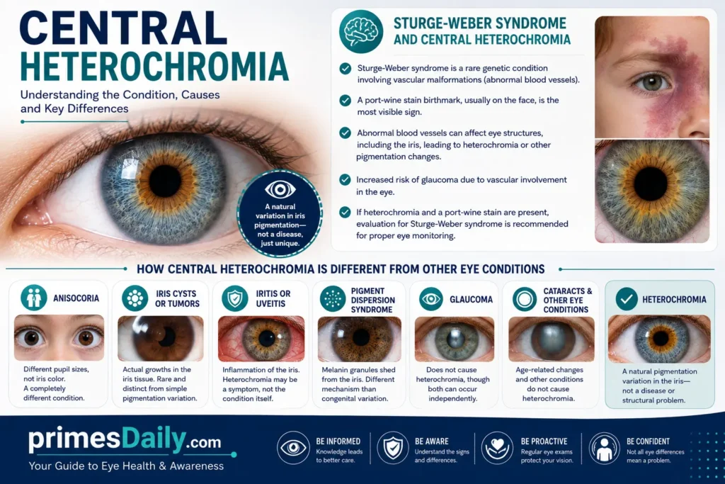
Sturge-Weber syndrome is a rare genetic condition involving vascular malformations (abnormal blood vessels).
The most visible sign of Sturge-Weber syndrome is a port-wine stain—a distinctive birthmark typically on the face. This birthmark is caused by abnormal blood vessels close to the skin surface.
The syndrome can also involve abnormal blood vessels affecting the eye structures, including the iris. This vascular involvement might result in heterochromia or other iris pigmentation changes.
People with Sturge-Weber syndrome involving the eye have increased risk of glaucoma due to the abnormal blood vessel involvement affecting eye structures.
If someone has heterochromia and a port-wine stain birthmark, evaluation for Sturge-Weber syndrome would be appropriate because the eye involvement needs careful monitoring.
How Central Heterochromia is Different From Other Eye Conditions
It’s worth clarifying how central heterochromia differs from various other eye conditions that might affect iris appearance or color.
Anisocoria is a condition where the pupils are different sizes. This is sometimes confused with heterochromia because an eye with an abnormally large or small pupil can appear different from the other eye. But anisocoria is about pupil size, not iris color. These are distinctly different conditions.
Iris cysts or tumors are actual growths in the iris tissue. These are rare but distinct from heterochromia, which is purely a pigmentation variation.
Iritis or uveitis are inflammation conditions affecting the iris. While inflammation can cause heterochromia, the heterochromia is a symptom of the inflammation, not the condition itself. Treating the inflammation treats the problem.
Pigment dispersion syndrome involves shedding of melanin granules from the iris. This can cause heterochromia or patchy iris appearance, but it’s a specific mechanism different from the melanin distribution variation of congenital central heterochromia.
Glaucoma does not cause heterochromia, though both conditions can occur in the same person independently.
Cataracts, age-related eye changes, and other vision conditions don’t cause heterochromia.
Heterochromia is purely a pigmentation variation in the iris—not a disease, not a structural problem, not a medical condition requiring treatment. It’s simply a difference in how melanin distributed.
Contact Lenses and Central Heterochromia
If you have central heterochromia and want to wear contact lenses, good news: you absolutely can.
Regular clear contact lenses work fine for people with central heterochromia. Your heterochromia won’t interfere with contact lens fitting or function. You might not even notice your heterochromia when wearing clear contacts since they’re virtually invisible.
If you want to wear colored contact lenses, you have that option too. Colored contacts can enhance your natural eye color, dramatically change your eye color, or even create cosmetic patterns.
Some people with central heterochromia choose colored contacts to hide their heterochromia if they’d prefer uniform eye color. Others enhance their heterochromia with colored contacts that complement their natural inner and outer ring colors.
The key is getting a proper contact lens fitting from an eye care professional. They’ll ensure the lenses fit correctly on your eye and that the prescription is appropriate if you need vision correction.
Contact lens care and hygiene are the same for everyone, including people with central heterochromia. Proper cleaning, storage, and replacement schedules keep your eyes healthy.
Some people worry that contacts might affect their heterochromia or that heterochromia might prevent them from wearing contacts. Neither is true. Contact lenses are safe and effective for people with central heterochromia.
Eye Care for People With Central Heterochromia
Taking care of your eyes when you have central heterochromia involves the same measures everyone should follow.
Regular eye exams help monitor your overall eye health. Even though central heterochromia doesn’t require specific monitoring, regular exams catch other potential eye conditions early. You should have comprehensive eye exams every one to two years, or more frequently if recommended by your eye care professional.
UV protection is important for everyone. Wear sunglasses with 100% UV protection to shield your eyes from sun damage. This matters whether or not you have heterochromia, but it’s worth emphasizing for anyone with distinctive eyes they want to keep looking their best.
Eye injury prevention protects your eyes. Wear appropriate protective eyewear when doing activities with injury risk. This prevents trauma that could cause acquired heterochromia or other serious eye injuries.
Screen time management and proper ergonomics reduce digital eye strain. Taking breaks from screens, positioning screens at appropriate distances, and maintaining proper lighting help prevent eye fatigue.
Maintaining overall health supports eye health. Eating a nutritious diet, exercising regularly, managing stress, and getting adequate sleep all support good eye health.
If you use contact lenses, proper care and hygiene are essential. Follow recommended replacement schedules and cleaning protocols to prevent infections.
If you use eye drops—whether for dry eyes or for conditions like glaucoma—use them as directed and follow up with your eye care professional about any side effects or concerns.
Monitoring Changes in Central Heterochromia
Most people with congenital central heterochromia never need to monitor their heterochromia because it’s stable and doesn’t change.
However, keeping track of your eye appearance over time is good practice. You might take occasional close-up photos of your eyes to document their appearance. If you ever notice changes, you’ll have previous photos to compare.
If you develop acquired heterochromia, monitoring for additional changes is important. The change that occurred might progress, stay stable, or even partially reverse depending on what caused it.
If you’re using prostaglandin analog eye drops for glaucoma, your ophthalmologist will monitor your eye health including iris changes. They’ll discuss any iris color changes that develop as a side effect.
Most people don’t need to do anything special to monitor their heterochromia. Your eye care professional will assess it during regular eye exams. If any concerns arise, they’ll discuss them with you.
When Central Heterochromia Requires Medical Evaluation
Most central heterochromia doesn’t require medical evaluation beyond what you’d get in a regular eye exam. But certain situations warrant specific assessment.
Sudden changes in iris color or development of new color variation warrants evaluation. Something caused that change, and determining what is important for your health.
Accompanying symptoms like eye pain, vision changes, redness, discharge, light sensitivity, or floaters suggest a problem requiring evaluation beyond simple heterochromia assessment.
Development of heterochromia in children should be evaluated to determine whether it’s congenital or acquired and to rule out underlying conditions.
Any heterochromia suspected to be related to a genetic syndrome should be evaluated for the complete syndrome.
Heterochromia accompanied by other symptoms like drooping eyelids, pupil size differences, or facial pigmentation changes warrants evaluation for related syndromes.
If you’re considering genetic counseling—perhaps because you’re concerned about inheritance or because heterochromia is part of a diagnosed genetic condition—that’s appropriate to discuss with your healthcare provider.
For most people with stable, congenital central heterochromia, a simple acknowledgment during a routine eye exam is all that’s needed.
Photography Tips for Central Heterochromia
If you have central heterochromia and enjoy photography, your distinctive eyes offer wonderful opportunities for striking self-portraits.
Lighting is crucial for capturing central heterochromia effectively. Natural light, especially from a window or outdoors in shade, reveals heterochromia beautifully. Direct sunlight can create glare that obscures the color details.
Close-up eye photography works best. Get close enough to fill the frame with your eye so the iris details are clearly visible. Macro lenses or the macro mode on phone cameras work well.
Look directly at the camera. Looking straight ahead shows your iris fully and captures the ring pattern most effectively.
Avoid heavy eye makeup that might obscure the iris details. Light makeup or no makeup photographs best if you want to showcase your heterochromia clearly.
Experiment with different angles. Slightly tilted angles sometimes reveal the color pattern better than straight-on views.
Camera settings matter. Use a fast shutter speed to avoid blur, proper focus on the iris, and adequate lighting to show the colors accurately.
Post-processing can enhance but shouldn’t drastically alter the natural colors. Slight adjustments to brightness or color saturation might make your heterochromia even more striking, but major alterations look artificial.
Sharing photos of your eyes with others might help raise awareness about central heterochromia and show others what it looks like.
Psychological and Social Aspects of Central Heterochromia
Having central heterochromia can carry psychological and social dimensions worth considering.
For many people, distinctive eyes become part of their identity and self-image in positive ways. They appreciate their unique appearance and incorporate their heterochromia into how they present themselves to the world.
Others might initially feel self-conscious about having different-looking eyes. During childhood and adolescence particularly, looking different from peers can feel uncomfortable.
Social reactions vary. Some people encounter positive comments and compliments about their unusual eyes. Others experience curiosity or even judgment about their appearance.
Building confidence in your appearance, including your distinctive eyes, contributes to overall self-esteem and wellbeing.
If you’re experiencing genuine distress about your appearance or heterochromia, speaking with a therapist or counselor can help. They can support you in developing positive self-image and coping strategies.
Most people eventually come to accept or even celebrate their unique features, including central heterochromia. What might initially seem unusual becomes simply “how my eyes look” and eventually becomes a valued part of your appearance.
Connecting with others who have central heterochromia, whether online or in person, can provide perspective and validation. Knowing others share your experience normalizes it.
Recent Research on Central Heterochromia (2024-2026)
Recent genetic research continues advancing our understanding of eye color determination and pigmentation variations like central heterochromia.
2024-2026 studies have focused on identifying additional genes involved in iris pigmentation and color variation. As genomic sequencing becomes more affordable and accessible, researchers can analyze more individuals with heterochromia, identifying genetic patterns.
Research on melanin distribution during fetal eye development is providing better understanding of the biological processes that create heterochromia. Imaging technologies allowing visualization of iris development in animal models are revealing mechanisms of melanin deposition.
Clinical studies examining the prevalence of different heterochromia types across various populations are helping document how common these variations actually are. Previous underdiagnosis of subtle heterochromia is being corrected as awareness improves.
Studies on prostaglandin analog side effects continue documenting the incidence and characteristics of medication-induced iris color changes. This helps ophthalmologists counsel patients about expected effects.
Research on inherited heterochromia patterns in families has provided better understanding of inheritance, confirming that most cases are not inherited despite being genetic.
Future research might enable earlier detection of acquired heterochromia associated with disease, potentially improving outcomes in conditions like glaucoma or uveitis where iris changes can signal problems.
Genetic Testing for Central Heterochromia
Do you need genetic testing if you have central heterochromia? For most people, the answer is no.
Genetic testing isn’t routinely recommended for congenital central heterochromia because it’s benign and identifying the specific genes involved doesn’t change management or treatment.
If your central heterochromia is part of a genetic syndrome (Waardenburg, Sturge-Weber, etc.), genetic testing for that syndrome might be appropriate. This helps with managing the overall condition and understanding inheritance patterns.
If you’re planning to have children and wondering whether you’ll pass heterochromia to them, genetic testing won’t change the answer since inheritance is unpredictable. Genetic counseling might be more useful than testing in this scenario.
If you’re experiencing acquired heterochromia or symptomatic heterochromia, the focus would be on determining the cause rather than genetic testing. Medical evaluation and imaging would be more relevant than genetic analysis.
If you have specific concerns about inheritance or genetic aspects of your heterochromia, discussing these with a genetic counselor or your physician can help determine whether testing is appropriate in your situation.
Central Heterochromia and Diabetes
Can diabetes cause heterochromia? Yes, diabetes can potentially cause acquired heterochromia as a rare complication.
Diabetes affects blood vessels throughout the body, including those in the eyes. Diabetic retinopathy, a complication affecting the retina from damaged blood vessels, is well-known. But diabetes can also affect other eye structures including the iris.
Changes in iris pigmentation or heterochromia can rarely occur as a manifestation of diabetic eye disease. This would be classified as acquired heterochromia resulting from the metabolic changes diabetes causes.
More commonly, people with diabetes should be concerned about diabetic retinopathy, diabetic macular edema, and increased risk of glaucoma and cataracts. These are more frequent complications than heterochromia.
If you have diabetes and notice iris color changes, discussing this with your ophthalmologist is important. It might represent diabetes-related eye changes requiring specific management.
Regular eye exams are crucial for people with diabetes to detect diabetic eye disease early. These exams would also reveal any heterochromia or iris changes.
Eye Surgery and Central Heterochromia
Can eye surgery cause central heterochromia? Yes, eye surgery can potentially cause acquired heterochromia as a rare complication.
Any surgery affecting the iris or affecting nerves controlling the iris could theoretically alter iris appearance or cause color changes. However, this is quite rare.
Cataract surgery, the most common eye surgery, doesn’t cause heterochromia. The surgery involves the lens, not the iris structures.
Glaucoma surgery affecting drainage structures might theoretically affect iris appearance, but this is exceptionally rare.
Complications from surgery—such as inflammation or iris tissue damage—could potentially cause acquired heterochromia, but this would be an unusual complication.
If you’re considering eye surgery, discussing this with your ophthalmologist can provide specific information about risks including very rare possibilities of iris changes.
Most people who have eye surgery never experience any iris color changes or heterochromia as a result.
Latanoprost and Central Heterochromia: Medication-Related Changes
Latanoprost, a prostaglandin analog eye drop used to treat glaucoma, is the most well-documented medication causing iris color changes.
How does latanoprost cause heterochromia? The medication stimulates melanin production in melanocytes (melanin-producing cells) in the iris. Over time, increased melanin deposition can darken the iris or create heterochromia if the color change is uneven.
The effect is dose-related and time-dependent. People using latanoprost for longer periods have higher likelihood of experiencing iris color changes. Some people show changes within months, others take years.
The changes are usually permanent or very slow to reverse even after discontinuing the medication. If you’ve been using latanoprost and develop iris color changes, those changes likely won’t return to baseline after stopping the medication.
Approximately 33% of people using latanoprost for 5 years or longer experience noticeable iris color changes. Most people experience slight darkening, but some develop actual heterochromia as the color change is uneven across the iris.
Is this dangerous? The iris color change itself isn’t dangerous or harmful to vision. It’s a cosmetic side effect, albeit a permanent one. This matters because you should know about it before starting the medication so you can decide if it’s acceptable to you.
If you’re using latanoprost and concerned about iris color changes, discussing this with your ophthalmologist is appropriate. They might discuss alternative glaucoma medications that don’t cause iris changes, though latanoprost’s effectiveness for glaucoma control must be weighed against its cosmetic side effects.
Glaucoma and Central Heterochromia
Does glaucoma cause heterochromia? Not directly. Glaucoma is a disease affecting eye pressure and the optic nerve. It doesn’t directly cause iris color changes.
However, certain glaucoma treatments—particularly prostaglandin analogs like latanoprost—can cause iris color changes including heterochromia as discussed above.
Some conditions causing both glaucoma and heterochromia exist (such as pigment dispersion syndrome or iris ectropion syndrome), but the heterochromia and glaucoma are separate manifestations of the same underlying condition.
If you have heterochromia and glaucoma, the heterochromia might be unrelated to the glaucoma. Alternatively, if both developed due to the same condition or if one resulted from treatment of the other, your ophthalmologist can explain the relationship.
Regular glaucoma screening is important regardless of whether you have heterochromia. Glaucoma is a serious condition requiring monitoring and treatment to prevent vision loss.
Inflammation of the Iris and Central Heterochromia
Inflammation of the iris (iritis) or the broader uveal tract (uveitis) can cause acquired heterochromia.
When the iris becomes inflamed, the inflammatory process can affect melanin-producing cells and melanin distribution. This can result in heterochromia or other iris color changes.
Iritis can have various causes including infection, autoimmune conditions, trauma, or sometimes unknown causes. Treating the underlying inflammation is important to prevent vision complications.
Chronic uveitis or repeated episodes of inflammation might result in permanent iris color changes or heterochromia even after inflammation resolves.
If you develop heterochromia along with eye redness, pain, or vision changes, evaluation for inflammatory causes would be appropriate.
Central Heterochromia Prognosis: What to Expect
For congenital central heterochromia, the prognosis is excellent. You have a lifetime condition that’s stable, permanent, and entirely benign.
You can expect your eye color pattern to remain the same throughout your life. The heterochromia won’t worsen, won’t cause complications, and won’t affect your vision or health.
Most people with central heterochromia live entirely normal lives, unaware that their distinctive eye color even has a medical term.
For acquired central heterochromia, the prognosis depends on what caused it. If it resulted from medication like latanoprost, you can expect it to be permanent. If it resulted from injury or inflammation, it depends on the underlying condition.
In terms of vision prognosis, heterochromia itself doesn’t affect your visual prognosis. You won’t develop vision problems because of heterochromia.
Overall life expectancy and general health are unaffected by central heterochromia. You have the same life expectancy and health prospects as anyone without heterochromia.
Will My Children Inherit Central Heterochromia?
This is a question many people with central heterochromia ask when planning families. The answer is probably not, but it’s not impossible.
Central heterochromia is usually not inherited. Even though it’s genetic (determined by your genes), you probably didn’t inherit it from your parents, and you probably won’t pass it to your children.
Most central heterochromia develops due to random genetic variation during fetal development. The specific melanin distribution pattern that created your heterochromia is unlikely to occur in your child’s development.
Theoretically, if the same random genetic variation occurs in your child, they might develop heterochromia. But this is not a predictable inheritance pattern.
If you have a family history of heterochromia—if multiple family members have it—then there might be a slightly increased likelihood in your children due to shared genetic factors. But even then, inheritance isn’t guaranteed.
If you’re planning to have children and are concerned about heterochromia inheritance, discussing this with a genetic counselor might provide peace of mind. But most people find that their children don’t develop heterochromia and that’s perfectly normal.
Age-Related Changes in Central Heterochromia
Do your eyes change as you age if you have central heterochromia?
The heterochromia pattern itself—the ring of different color around your pupil—remains stable throughout your life. The same ring that was present at birth will be present at age 80.
Other age-related eye changes affect everyone, including people with central heterochromia. Presbyopia (difficulty focusing on close objects) develops in your 40s and 50s. The lens becomes less flexible with age. The pupil dilates less in dim light. Vision naturally declines slightly with age.
But these age-related changes affect everyone equally regardless of heterochromia. They’re not caused by heterochromia and aren’t more severe in people with heterochromia.
Cataracts and age-related macular degeneration are eye conditions more common with age. These occur at similar rates in people with heterochromia and people without.
The color appearance of your heterochromia might change slightly with age if overall iris coloration changes, but the basic pattern remains the same.
By maintaining regular eye exams and managing any age-related eye conditions, people with central heterochromia can maintain good vision throughout their lives just like anyone else.
Central Heterochromia Documentation and Medical Records
Should you document your central heterochromia in medical records? It depends on the circumstances.
If your heterochromia is congenital and stable, simple documentation in your medical record noting its presence is sufficient. This helps future healthcare providers recognize the feature if they encounter it.
If your heterochromia is acquired or if it’s part of a genetic syndrome, more detailed documentation including dates of onset and associated conditions is appropriate.
Photography can be useful for documentation, especially if you want to track changes over time or if acquired heterochromia might progress.
If you have a genetic syndrome involving heterochromia, documentation of that diagnosis is crucial for managing the overall condition.
For routine congenital central heterochromia with no other complications, extensive documentation probably isn’t necessary beyond simple acknowledgment in your medical record.
Support Resources for Central Heterochromia
While central heterochromia isn’t a disease requiring treatment, support and information resources can be helpful for understanding your condition and connecting with others.
Your ophthalmologist or optometrist is your primary resource for information and guidance about your heterochromia. They can answer questions and provide reassurance.
Online support groups and forums connecting people with various eye conditions often include people with heterochromia sharing experiences and information.
Educational resources about eye health and eye conditions can provide scientific information about heterochromia.
Genetic counselors can provide guidance if you have questions about inheritance or if heterochromia is part of a genetic syndrome.
Mental health professionals can help if you’re experiencing psychological distress related to your appearance or heterochromia.
Family and friends providing support and acceptance helps build confidence and positive self-image.
FAQs
Is central heterochromia permanent? Yes, congenital central heterochromia is permanent and stable throughout your life. Acquired heterochromia might be permanent or gradual depending on its cause.
Can central heterochromia appear in one eye only? Yes, absolutely. You can have the ring pattern in just one eye while the other eye displays a single, solid color.
What’s the difference between central heterochromia and hazel eyes? Central heterochromia has a distinct ring pattern with clear color boundaries, while hazel eyes have blended colors throughout the iris.
Do I need to see a doctor about my central heterochromia? No, not specifically for the heterochromia if it’s congenital and stable. Regular eye exams are recommended for everyone for general eye health.
Will my central heterochromia affect my ability to wear contact lenses? No, you can wear regular or colored contact lenses if you have central heterochromia. The heterochromia doesn’t interfere with contact lens use.
Is central heterochromia genetic or inherited? Central heterochromia is genetic but usually not inherited. It’s determined by your genes but usually isn’t passed from parents to children.
Does central heterochromia affect my vision? No, heterochromia is purely cosmetic and doesn’t affect your vision or eyesight.
Can central heterochromia develop later in life? Yes, acquired heterochromia can develop due to eye injury, infection, inflammation, or medication. This warrants professional evaluation.
What percentage of people have central heterochromia? Exact percentages are unclear because central heterochromia is underdiagnosed, but it’s probably less common than 1% of the population.
Is central heterochromia dangerous? Congenital central heterochromia is not dangerous. Acquired heterochromia warrants investigation to determine its cause, but the heterochromia itself isn’t dangerous.
Can I hide my central heterochromia? Yes, you can wear colored contact lenses to obscure the heterochromia if you prefer uniform eye color.
Does heterochromia run in families? Not usually. Most people with central heterochromia didn’t inherit it from family members, though rare familial patterns exist.
Do people with central heterochromia have better or worse vision? No, vision is unrelated to central heterochromia. People with heterochromia have the same range of vision abilities as anyone else.
Is central heterochromia more common in certain ethnicities? Central heterochromia appears in all ethnic groups, though it might be more noticeable in people with lighter eye colors.
Can heterochromia be surgically corrected? No surgery can change your heterochromia. Colored contacts are the only practical way to alter its appearance if desired.
Conclusion
Central heterochromia is a fascinating natural variation in eye color where the iris displays two distinct colors in a ring pattern—a different shade around the pupil and another shade toward the outer iris edge.
For most people, central heterochromia is a benign, stable, lifelong cosmetic feature. It doesn’t affect vision, doesn’t indicate disease, and requires no medical treatment. It’s simply how your eyes happened to look due to genetics and chance.
Understanding what central heterochromia is, why it happens, and that it’s harmless can bring peace of mind if you have the condition yourself or know someone who does. If you have heterochromia that developed suddenly after birth, professional evaluation is worthwhile to determine its cause. But stable, lifelong heterochromia is nothing to concern yourself about.
Whether you view your central heterochromia as a distinctive feature to celebrate, a cosmetic variation to camouflage with contact lenses, or simply the way your eyes happen to look, understanding the biology behind it enriches your appreciation for the complexity and variation in human appearance. Your eyes are unique, just like you are.
For the latest, updated, and unique information, visit Puerto Rican Food.